


Medición del espacio tibiofemoral
Resumen
Fundamento: : la evaluación del estado actual y de progresión de la gonartrosis necesita de métodos tanto clínicos como imagenológicos, estos últimos necesitan de entrenamiento, mayor tiempo y familiarización por parte del personal médico con la tecnología digital.
Objetivo: mostrar dos métodos de medición del espacio articular tibiofemoral en pacientes con gonartrosis.
Métodos: la búsqueda y análisis de la información se realizó en un periodo de 56 días (primero de noviembre de 2018 al 26 de diciembre de 2018) y se emplearon las siguientes palabras: tibiofemoral space measure AND knee osteoarthritis, knee joint space narrowing AND knee osteoarthritis, knee joint space width a partir de la información obtenida se realizó una revisión bibliográfica de un total de 272 artículos publicados en las bases de datos PubMed, Hinari, SciELO y Medline mediante el gestor de búsqueda y administrador de referencias EndNote, de ellos, se utilizaron 31 citas seleccionadas para realizar la revisión, 27 de los últimos cinco años.
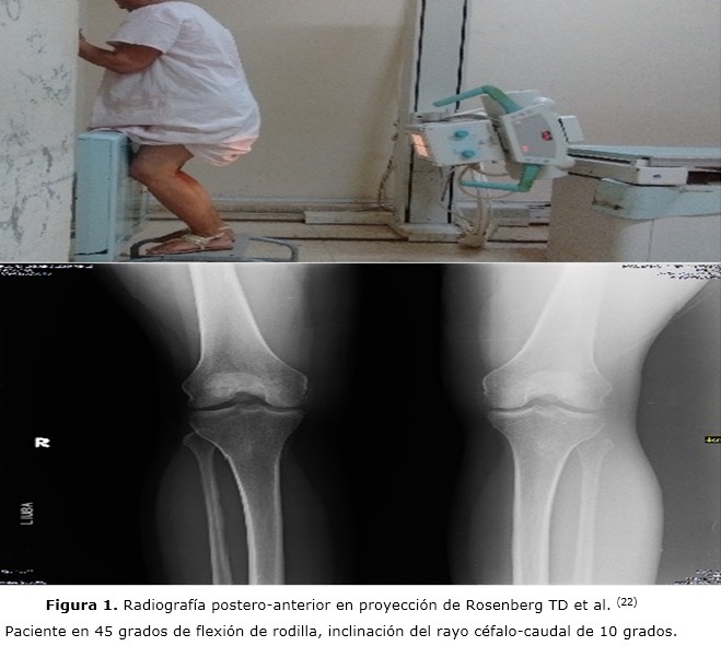
Resultados: se mencionan los métodos aplicables tanto en películas radiográficas convencionales y las de tipo digital. Se hace referencia a la vista radiográfica para la evaluación, gradación e interpretación del espacio articular tibiofemoral. Se exponen dos métodos de medición por pasos.
Conclusiones: la medición del espacio tibiofemoral es importante para conocer el estado en que se encuentran los enfermos que padecen de gonartrosis, así como, determinar la respuesta desde el punto de vista imagenológico a diferentes modalidades de tratamiento aplicados tanto conservadores como quirúrgicos.
DeCS: OSTEOARTRITIS DE LA RODILLA/diagnóstico por imagen; OSTEOARTRITIS DE LA RODILLA/cirugía; FEMUR/ anatomía&histologia; TIBIA/anatomía&histologia; INTENSIFICACION DE IMAGEN RADIOGRAFICA.
Palabras clave
Referencias
Eymard F, Parsons C, Edwards MH, Petit-Dop F, Reginster JY, Bruyère O, et al. Statin use and knee osteoarthritis progression: results from a post-hoc analysis of the SEKOIA trial. Joint Bone Spine. 2018 Oct;85(5):609-14.
Fryzowicz A, Dworak LB, Koczewski P. Prophylaxis of medial compartment gonarthrosis in varus knee current state of knowledge. Arch Med Sci. 2018 Mar;14(2):454-9.
Kemnitz J, Wirth W, Eckstein F, Culvenor AG. The role of thigh muscle and adipose tissue in knee osteoarthritis progression in women: data from the Osteoarthritis Initiative. Osteoarthritis Cartilage. 2018 Sep;26(9):1190-5.
Sgroi M, Faschingbauer M, Reichel H, Kappe T. Can the frontal tibiofemoral alignment be assessed on anteroposterior knee radiographs? J Orthop Traumatol. 2016 Dec;17(4):339-43.
Bloecker K, Wirth W, Guermazi A, Hitzl W, Hunter DJ, Eckstein F. Longitudinal change in quantitative meniscus measurements in knee osteoarthritis-data from the Osteoarthritis Initiative. Eur Radiol. 2015 Oct;25(10):2960-8.
Wirth W, Hunter DJ, Nevitt MC, Sharma L, Kwoh CK, Ladel C, et al. Predictive and concurrent validity of cartilage thickness change as a marker of knee osteoarthritis progression: data from the Osteoarthritis Initiative. Osteoarthritis Cartilage. 2017 Dec;25(12):2063-71.
Hafezi-Nejad N, Demehri S, Guermazi A, Carrino JA. Osteoarthritis year in review 2017: updates on imaging advancements. Osteoarthritis Cartilage. 2018 Mar;26(3):341-9.
Konin GP, Walz DM. Arthropathies, osteonecrosis, and bursitis. En: Scott WN, editor. Insall & Scott Surgery of the Knee. 5th ed. Philadelphia: Elsevier; 2012.p.e9-11.
Ilhanli İ, Güder N, Tosun A, Avci E, Çelik C. Computer-assisted joint space area measurement: a new technique in patients with knee osteoarthritis. Arch Rheumatol. 2017 Apr;32(4):339-46.
Culvenor AG, Wirth W, Ruhdorfer A, Eckstein F. Thigh muscle strength predicts knee replacement risk independent of radiographic disease and pain in women: data from the osteoarthritis initiative. Arthritis Rheumatol. 2016 May;68(5):1145-55.
Dell'isola A, Wirth W, Steultjens M, Eckstein F, Culvenor AG. Knee extensor muscle weakness and radiographic knee osteoarthritis progression. Acta Orthop. 2018 Aug;89(4):406-11.
Eymard F, Parsons C, Edwards MH, Petit-Dop F, Reginster JY, Bruyère O, et al. Diabetes is a risk factor for knee osteoarthritis progression. Osteoarthritis Cartilage. 2015 Jun;23(6):851-9.
Kellgram JH, Lawrence JS. Radiological assessment of ostoearthrosis. Ann Rheum Dis. 1957 Dec;16(4):494-502.
Clément J, Toliopoulos P, Hagemeister N, Desmeules F, Fuentes A, Vendittoli PA. Healthy 3D knee kinematics during gait: differences between women and men, and correlation with x-ray alignment. Gait Posture. 2018 Jul;64:198-204.
Cromer MS, Bourne RM, Fransen M, Fulton R, Wang SC. Responsiveness of quantitative cartilage measures over one year in knee osteoarthritis: comparison of radiography and MRI assessments. J Magn Reson Imaging. 2014 Jan;39(1):103-9.
Jamali AA, Meehan JP, Moroski NM, Anderson MJ, Lamba R, Parise C. Do small changes in rotation affect measurements of lower extremity limb alignment? J Orthop Surg Res. 2017 May;12(1):77.
Kan H, Arai Y, Kobayashi M, Nakagawa S, Inoue H, Hino M, et al. Fixed-flexion view X-ray of the knee superior in detection and follow-up of knee osteoarthritis. Medicine (Baltimore). 2017 Dec;96(49):e9126.
Ijaz Khan H, Chou L, Aitken D, McBride A, Ding C, Blizzard L, et al. Correlation between changes in global knee structures assessed by magnetic resonance imaging and radiographic osteoarthritis changes over ten years in a midlife cohort. Arthritis Care Res (Hoboken). 2016 Jul;68(7):958-64.
Ratzlaff C, Ashbeck EL, Guermazi A, Roemer FW, Duryea J, Kwoh CK. A quantitative metric for knee osteoarthritis: reference values of joint space loss. Osteoarthritis Cartilage. 2018 Sep;26(9):1215-24.
Parent ME, Vézina F, Carrier N, Masetto A. Indications for and clinical procedures resulting from magnetic resonance imaging of the knee in older patients: are we choosing wisely? Can Fam Physician. 2018 Mar;64(3):e126-e132.
Zhang M, Driban JB, Price LL, Harper D, Lo GH, Miller E, et al. Development of a rapid knee cartilage damage quantification method using magnetic resonance images. BMC Musculoskelet Disord. 2014 Aug;15:264.
Rosenberg TD, Paulos LE, Parker RD, Coward DB, Scott SM. The forty five degree posteroanterior flexion weight bearing radiograph of the knee. J Bone Joint Surg Am. 1988 Dec;70(10):1479-82.
Huang TL, Wang CC, Yang KC, Wu CC. Reliability of roentgenographic knee alignment measurements in gonarthrosis. J Knee Surg. 2018 Apr;31(4):302-5.
Pinsornsak P, Naratrikun K, Kanitnate S, Sangkomkamhang T. The one-leg standing radiograph: an improved technique to evaluate the severity of knee osteoarthritis. Bone Joint Res. 2016 Sep;5(9):436-41.
Rueckl K, Boettner F, Maza N, Runer A, Bechler U, Sculco P. The posterior-anterior flexed view is better than the anterior-posterior view for assessing osteoarthritis of the knee. Skeletal Radiol. 2018 Apr;47(4):511-517.
Mehta N, Duryea J, Badger GJ, Akelman MR, Jones MH, Spindler KP, et al. Comparison of 2 radiographic techniques for measurement of tibiofemoral joint space width. Orthop J Sports Med. 2017 Sep;5(9):1-9.
Halilaj E, Le Y, Hicks JL, Hastie TJ, Delp SL. Modeling and predicting osteoarthritis progression: data from the osteoarthritis initiative. Osteoarthritis Cartilage. 2018 Dec;26(12):1643-50.
Hunter DJ, Le Graverand MP, Eckstein F. Radiologic markers of osteoarthritis progression. Curr Opin Rheumatol. 2009 Mar;21(2):93-4.
Komatsu D, Hasegawa Y, Kojima T, Seki T, Higuchi Y, Ishiguro N. Absence of a relationship between joint space narrowing and osteophyte formation in early knee osteoarthritis among Japanese community-dwelling elderly individuals: a cross-sectional study. Mod Rheumatol. 2017 Jul;27(4):675-82.
Shon OJ, Park SJ, Shim BJ, Lee DY. Comparative study of clinical and radiographic outcomes of high tibial osteotomy in patients with kissing lesions and non-kissing lesions. Knee Surg Relat Res. 2017 Dec;29(4):288-94.
Mezlini-Gharsallah H, Youssef R, Uk S, Laredo JD, Chappard C. Three-dimensional mapping of the joint space for the diagnosis of knee osteoarthritis based on high resolution computed tomography: comparison with radiographic, outerbridge, and meniscal classifications. J Orthop Res. 2018 Sep;36(9):2380-91.
Enlaces refback
- No hay ningún enlace refback.

Esta obra está bajo una licencia de Creative Commons Reconocimiento-NoComercial-CompartirIgual 4.0 Internacional.
 La Revista está: Certificada por el CITMA
 Acreditados como: "Web de Interés Sanitario"